


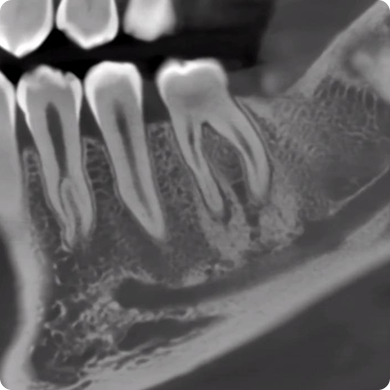
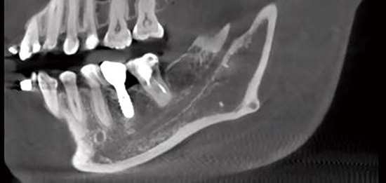
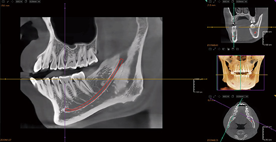
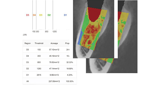
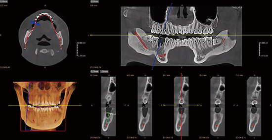
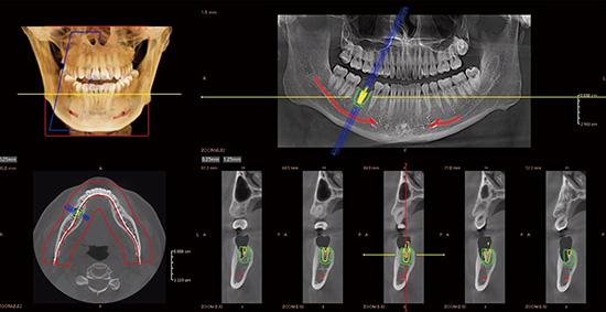
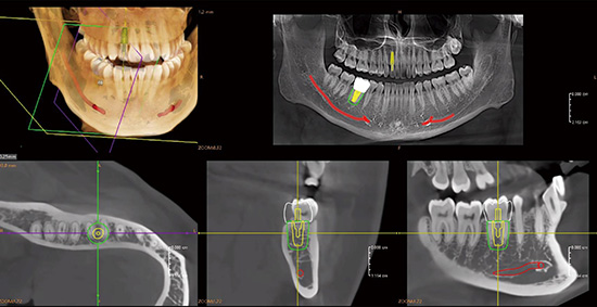
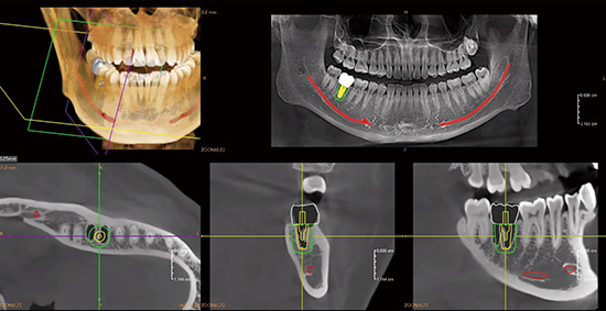
Single-Tooth Implant Planning
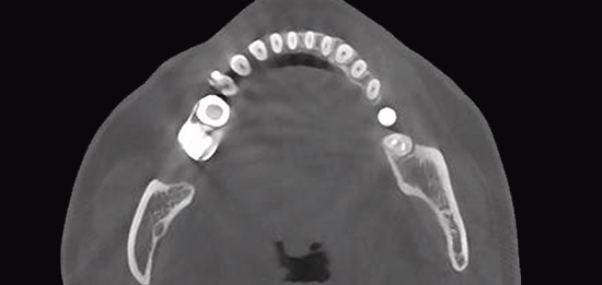
Full-Arch Implant Planning
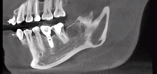
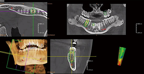
Implant Placement with GBR
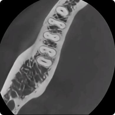
TMJ
Airway Analysis
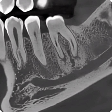
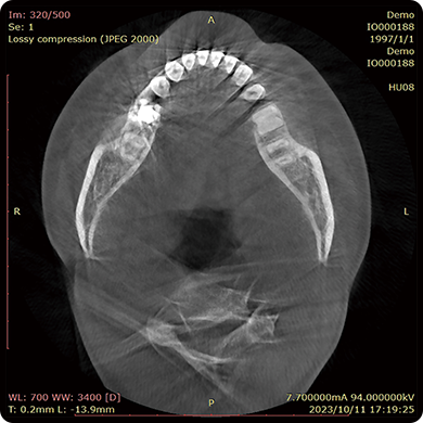
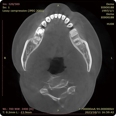
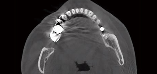
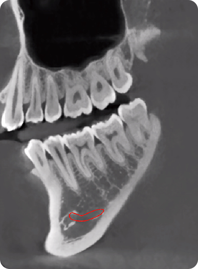
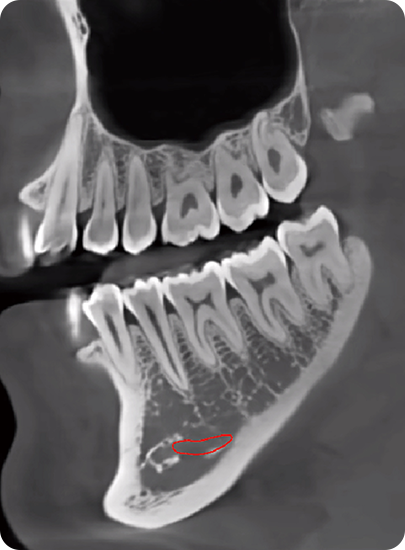
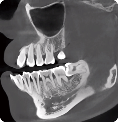
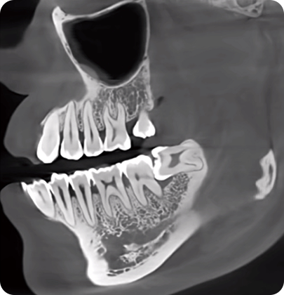
Oral Health Examination
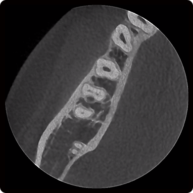
Maxillary Sinus Diagnosis
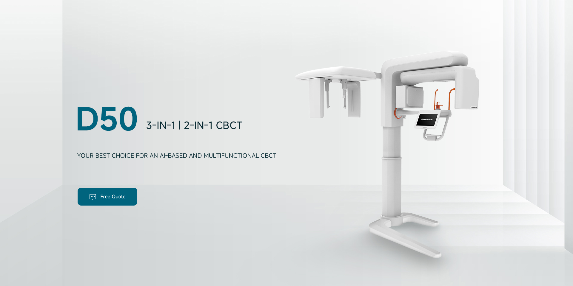


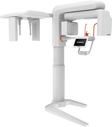
3-IN-1
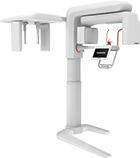
| 2-IN-1
|
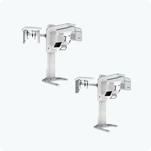
CBCT |
PANO |
CEPH | |
| D50 2 in 1 |  | | - |
D50 3 in 1 | | | |
| SPECIFICATIONS | PARAMETER |
|---|---|
| Tube Voltage | 60~90 KV |
| Tube Current | 4~10 mA |
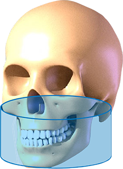
| FOV(Diameter*Height) | 15*9; 5*5 |
| Focal Spot | 0.5mm |
| Gray Scale | 16 bit |
| Voxel | 0.25; 0.2; 0.1; 0.075mm |
| Total Filtration | 2.5mm Al |
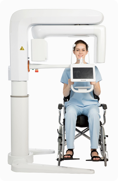
| Scan Position | Standing and Wheelchair |
| Exposure Time | ≤15s |
| Reconstruction Time | <45s |
| Machine Dimension |
2 in 1 3 in 1 |
1650(L)*1060(W)*1733~2283(H)mm 1650(L)*1840(W)*1733~2283(H)mm |
| Recommended installation Space |
2 in 1 3 in 1 |
1900(L)*1300(W)*2300(H)mm 1900(L)*2100(W)*2300(H)mm |