
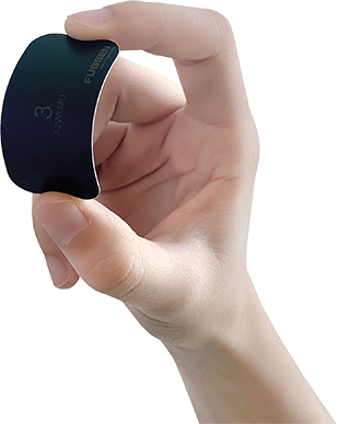
Imaging Plate Size:
0,1,2,3
Theoretical Resolution:
12Lp/mm
USB Interface:
USB2.0, USB3.0
Size(H*W*D):
260×167×325mm
Theoretical Gray Scale:
14bit
Weight
5.2kg
Theoretical Pixel Size
42μm
Data Format
DICOM3.0
Photophor Records
Auto
Power Supply
100-240V(AC),50/60Hz,1.5A
Operating System
Windows 7, Windows 8, Windows 10
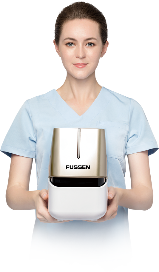
Imaging Plate Scanner · F200
Digital Intraoral Imaging Plate Scanning System
Simple and Convenient Operation
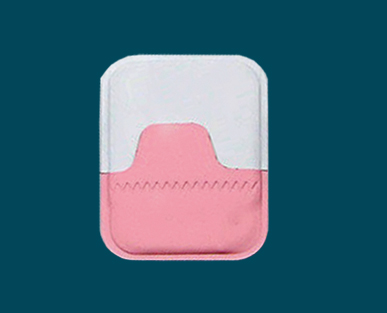
Four size of imaging plate
satisfies various clinical applicationOnly 0.1mm thickness,
Ultrathin designBendng at 270 degree
for more comfortable ixperienceOver 2000
times use
Four size of imaging plate
satisfies various clinical applicationOnly 0.1mm thickness,
Ultrathin designBendng at 270 degree
for more comfortable ixperienceOver 2000
times use

Operation method
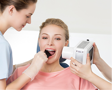 1. Position the patient and place the phosphor film for shooting
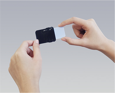 2. Take out the imaging plate
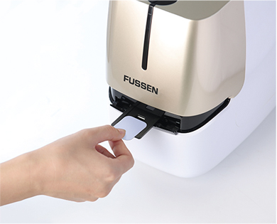 3. Touch the button on top of the device to acquire image
Auto Erace
Automatically erase latent image information
on phosphor while image acquisition,
convenient for next capturing

Compact Design
The overall unit adopt compact design,
portable and save space.

HD Image Display
Up to 42 μm ultra high resolution, clearly distinguish enamel,
root tip, periodontal ligament, pulp cavity and other fine anatomical structures.
Assist doctor to make accurate diagnosis by the high quality clinical images.

Auto Erace
Automatically erase latent image information
on phosphor while image acquisition,
convenient for next capturing
Compact Design
The overall unit adopt compact design,
portable and save space.
HD Image Display
Up to 35 μm ultra high resolution, clearly distinguish enamel,
root tip, periodontal ligament, pulp cavity and other fine anatomical structures.
Assist doctor to make accurate diagnosis by the high quality clinical images.
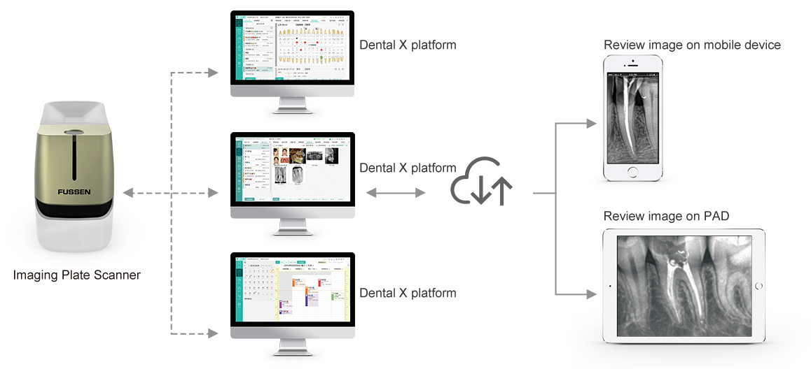
Review Image On Mobile Devie
Wireless transmission makes it easy to review images whenever and wherever possible
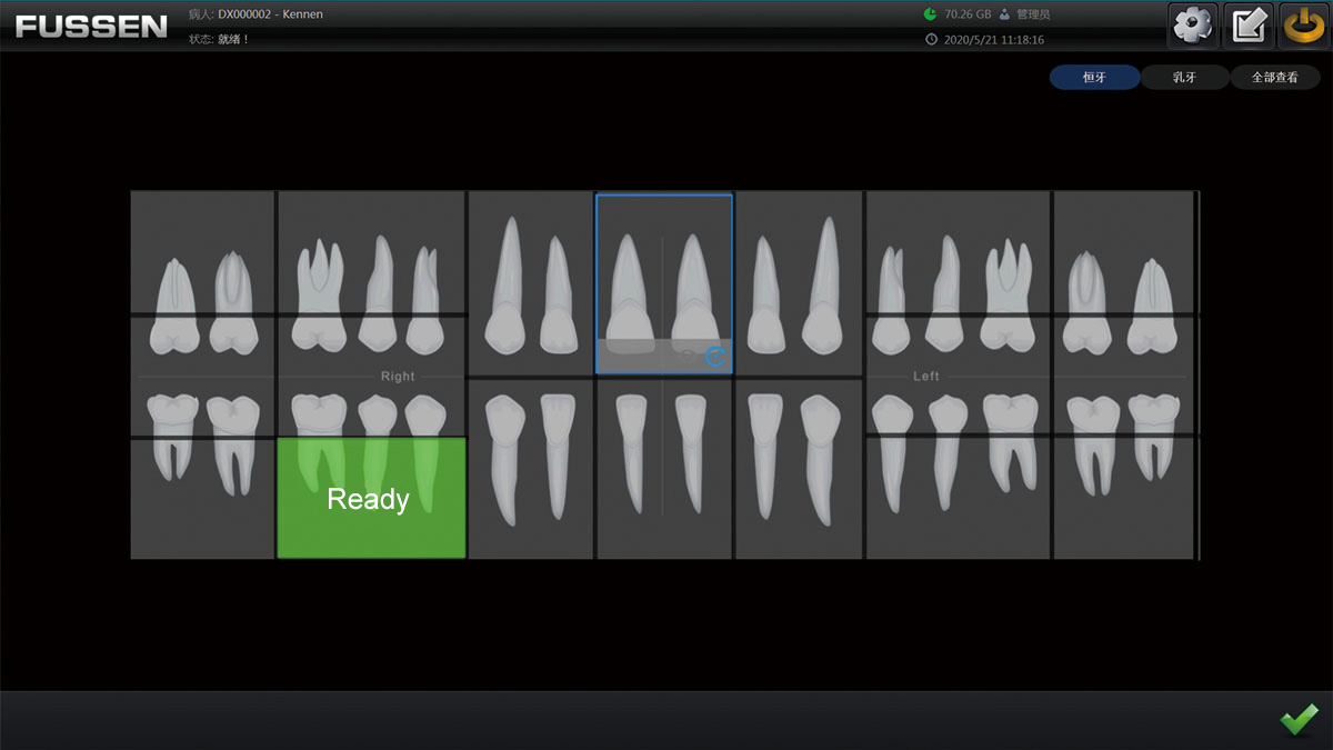
Friendly Operation Interface
Succinct and high-efficient operation interface, bring you operating comfortably

HD Images More in line with your demand
Up to 42 μm resolution, clearly distinguish root tip, periodontal ligament, trabecular bone and other fine anatomical structures.
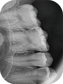 Maxillary molars
 Maxillary anterior teeth
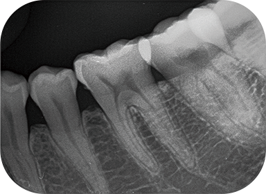 Mandibular premolars
Clinical Application
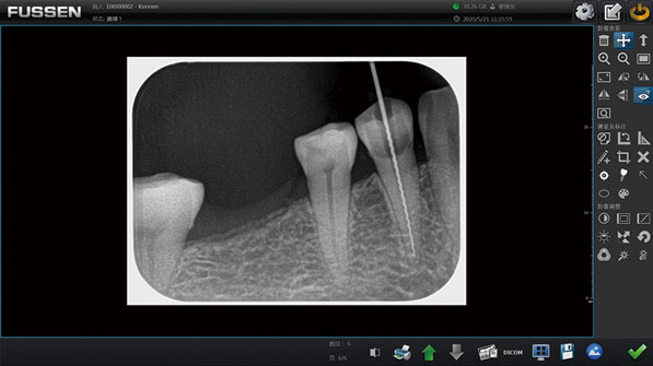
Acquire high quality clinical images, assist doctors to make accurate diagnostis
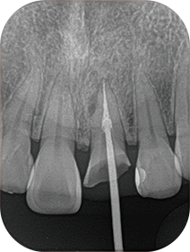 Endodontic treatment
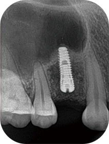 Healing check after implant surgery
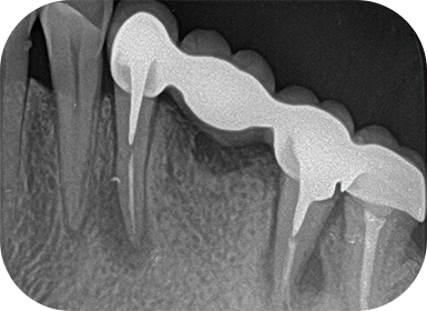 Periapical treatment
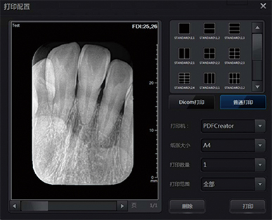
Data Print and Export Format
Support DICOM printer and general printer
including DICOM, DICOMDIR, BPM and JPG
Difference From Traditional Film
Fast imaging, reduce waiting time, Avoid photographic developing and fixing Non chemical storage and avoid environment pollution impact Permanent storage of digital images, be helpful to comparison before treatment High definition, not affected by exposure conditions and radiography techniques

14 Gray Scale Imaging
Compared with 12 gray scale, 12288 gray level increased
The image is more delicate and clear