Tube Voltage:
60~90 KV
Tube Current:
4~10 mA
FOV (Diameter * Height):
15*9,5*5 cm
Total filtration:
2.5 mmAl
Focal Spot:
0.5 mm
Voxel:
0.25, 0.2, 0.1, 0.075 mm
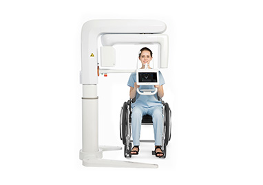
Scan Posture:
Standing
Type of X ray:
Pulse
Gray Scale:
14 bit
Scan Time:
≤15 s
Reconstruction Time:
≤60 s
Machine Dimension:
1560(L) * 1960(W) * 1733~2283(H) mm
Recommend Installation Space:
1900(L) * 2100(W) * 2300(H) mm
Medium to Large CBCT · D50
FOV 15*9- Greater Vision
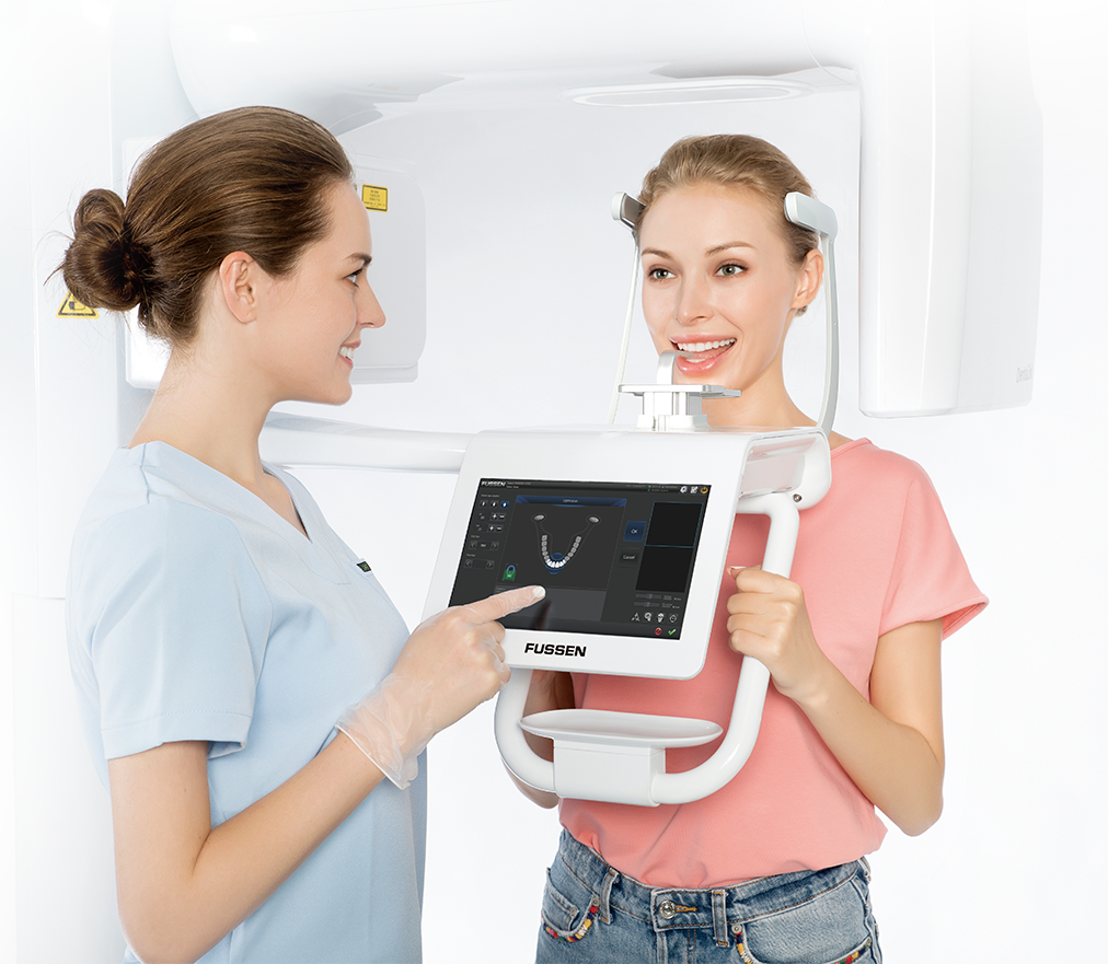
Design for patient experience - High performance with modest price
More practical product performance for clinical use
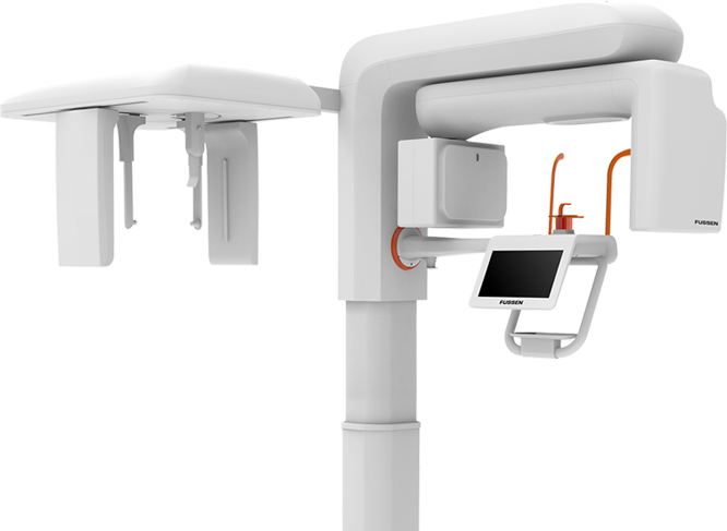
3 in 1 CBCT More in line with your demand
D55 3 in 1 CBCT includes CT, Panaramic Imaging and Cephalometric. By combining image processing and Fussen's R&D experience, a new software algorithm is introduced to clearly display the anatomical structures such as whole dentition, maxilla and mandible, zygoma, superior frontal sinus, mandibular nerve canal, periapical tissue and periodontal ligament, which will improve the accuracy of diagnosis and treatment, increase the convenience of treatment plan and patient satisfaction.

Pano
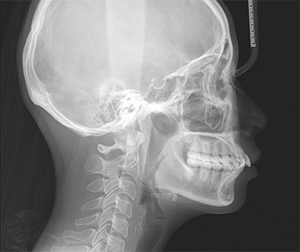
Ceph

CBCT
Usable FOV

FOV 5cm×5cm
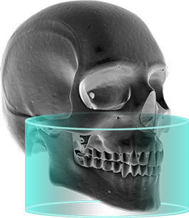
FOV 15cm×9cm
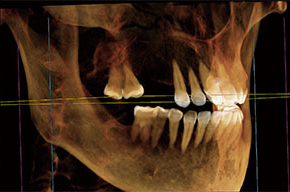
MARS Anti-artifacts technology
D55 breaks the traditional dental imaging convention, adopts the new Mars technology to remove metal artifacts, effectively reduces the metal artifacts. Check surrounding conditions of the implant and implant bone combination clearly. The image has more diagnostic value.
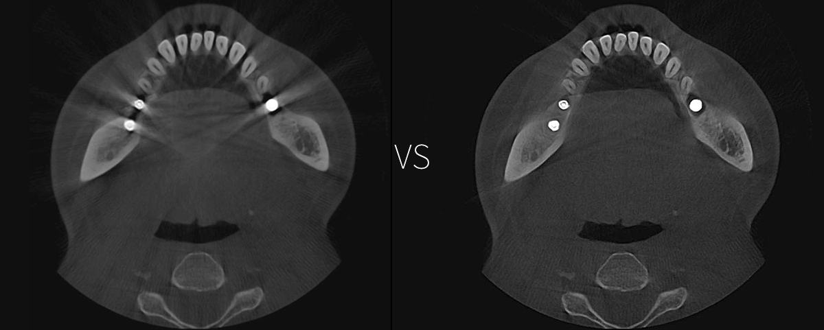
FOV 15cm×9cm
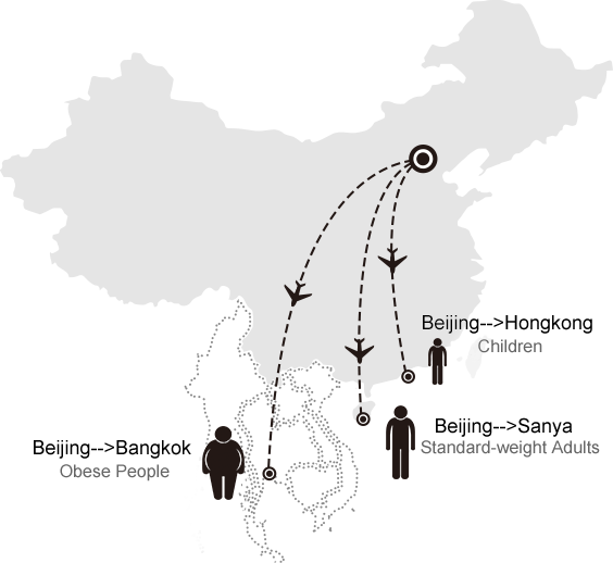
Super Low Radiation Dose
Radiation dose of one CBCT exam ≈ 3 hours flight
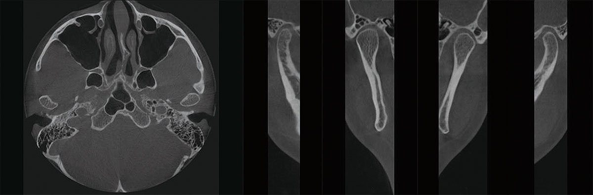
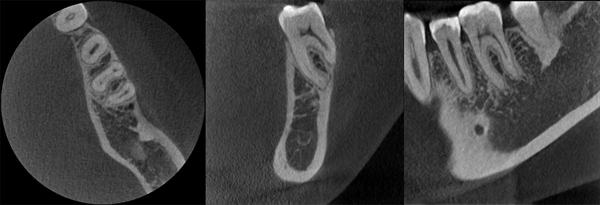
Authentic and Reliable Image particulars

Posterior alveolar artery of
lateral wall of maxillary sinus
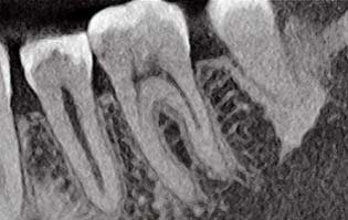
Root tip
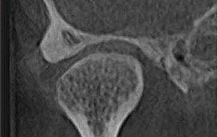
TMJ
Mandibular canal
Extreme Panoramic, Focusing On Image particulars

Panoramic Image of Patient
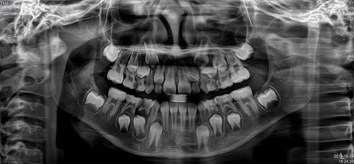
Panoramic Image of Child
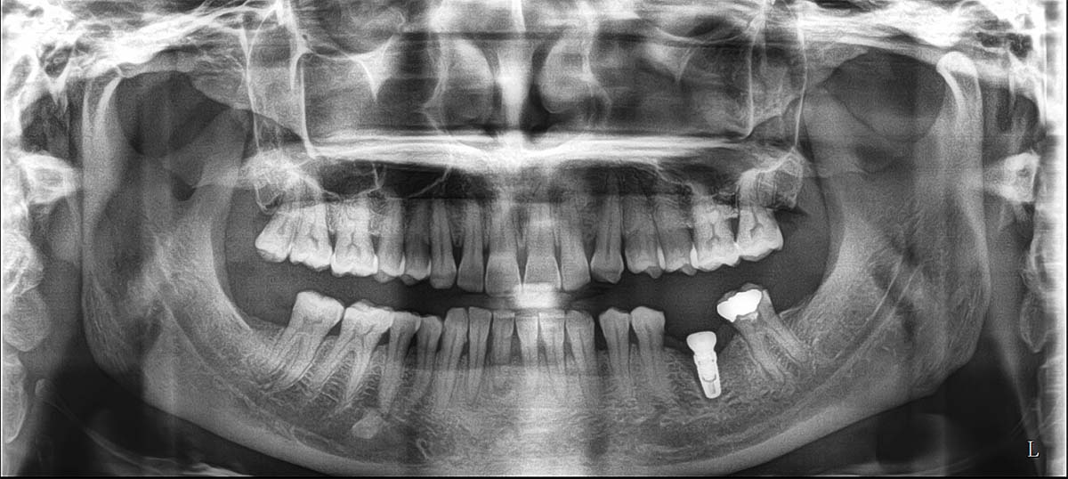
Panoramic Image of Dental Implant
Dental X - The profession digital image management and treatment plan online map out platform
Both Sides TMJ
-15*15 big FOV to facilitate doctor to diagnostis TMJ disease
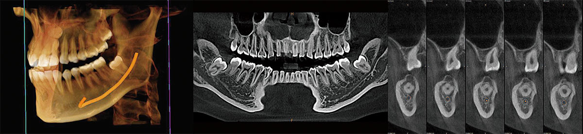
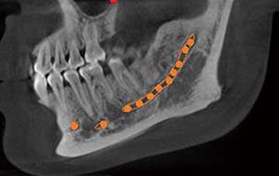
Outline Mandibular Canal
-View the path of mandibular canal, position relation with wisdom tooth
-View the distance between mandibular canal and implant, avoid implant touching the canal
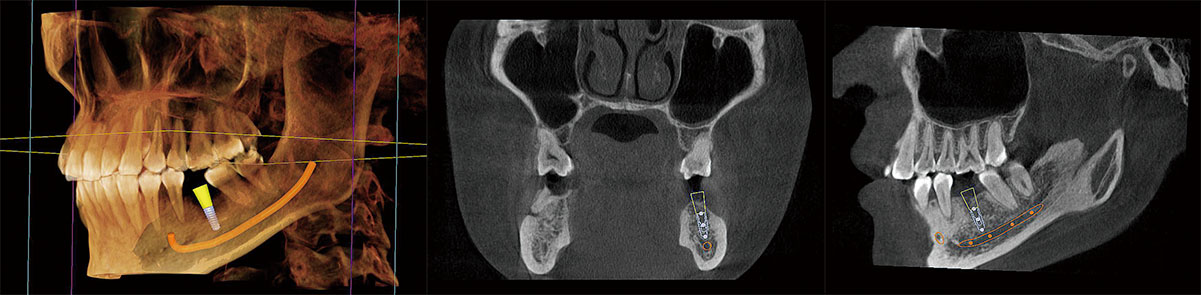
Dental Implant Simulation
-DViewMatrix adds new 3D dental implant simulation module to measure width, height and density of alveolar bone of the missed tooth, and outline mandibular canal
-Abundant implant bank
-Design scheme of simulated dental implant by inputting implant parameters
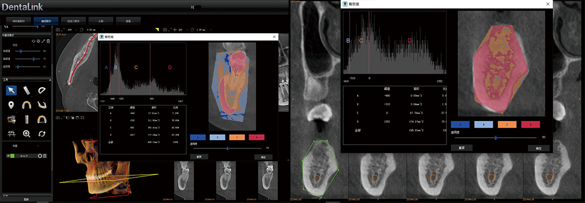
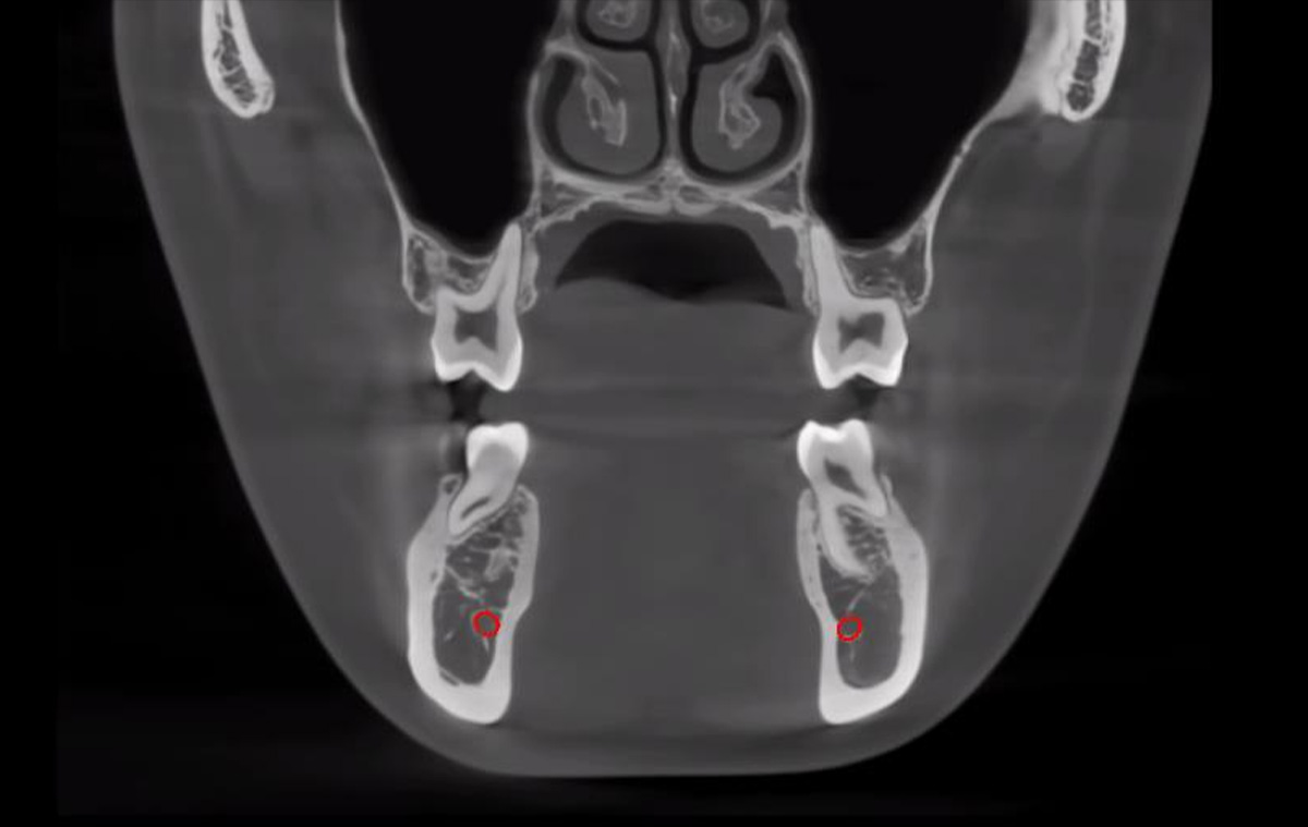
Bone Density Measurement
Check the bone density to make decision
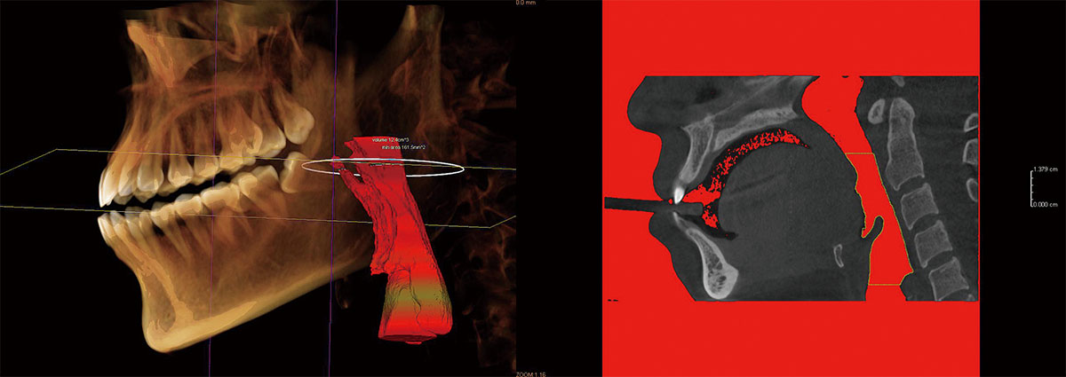
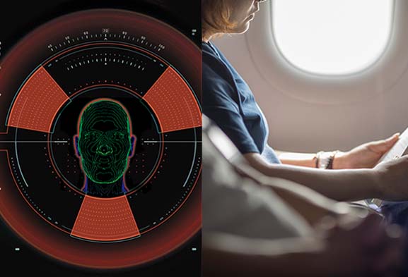
Airway Analysis
-Airway analysis data is necessary for orthodontics treatment planning, for airway volume measurement of any region, and for automatic determination of airway cross-sectional area at the narrowest position
-It is more important for snoring disease or OSAS patient. Doctors may design treatment through airway analysis
More Valuable Clinical Application
Maxillary Sinus Diagnosis
Mandibular Single Tooth Implant
Examination (Pre-surgery)

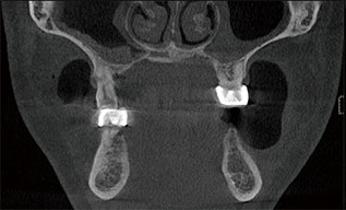
Mandibular Single Tooth Implant
Examination (Post-surgery)
Maxillary Multiple Teeth Implant
Examination (Pre-surgery)

Maxillary Multiple Teeth Implant
Examination (Post-surgery)
Root Canal Mode

Full Dentition Examination
C Type Root Canal
More Valuable Clinical Application

Low dose mode (ALARA mode, Root canal mode, Child mode)
User friendly handrill and tray design, to ease patient